
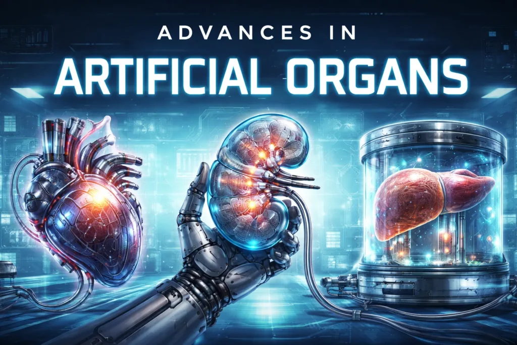
Imagine a world where “organ transplant” doesn’t mean waiting by the phone for a miracle, hoping a stranger’s family says yes in their moment of grief. Imagine a world where we don’t have to rely on animal testing to develop new drugs, because we can test them on tiny, living replicas of human organs instead. And imagine a world where repairing a failing heart or a damaged kidney is as routine as getting a flat tire fixed at a garage—using parts that are custom-made just for you.
This isn’t the plot of a sci-fi movie. This is the real-life revolution happening right now in labs all over the world. We are living through a golden age of discovery in artificial organs. Scientists, engineers, and doctors are teaming up to do the impossible: they are learning to build our bodies’ parts, piece by piece, cell by cell. They’re using tools like 3D printers, stem cells, and even tiny computer chips to create the future of medicine.
Let’s dive into this incredible story and meet the heroes—both human and technological—that are making it all possible. This journey will take us from the smallest building blocks of life to the most advanced machines ever created, and along the way, we’ll discover how close we really are to a world where no one dies waiting for a transplant.
The Long Wait: A Problem of Life and Death
To understand why this work is so urgent, we have to understand the problem. Right now, there are hundreds of thousands of people around the world waiting for an organ transplant. In the United States alone, over 100,000 people are on the national waiting list, most of them needing a kidney. They are stuck in a terrible race against time, hoping a donor will be found before their body gives out. Around 14% of American adults have chronic kidney disease, a trend that’s only getting worse, which means the demand for healthy organs keeps growing while the supply stays painfully limited.
Every single day, about seventeen people die waiting for an organ in the United States. That’s seventeen families getting devastating news, seventeen stories cut short, all because there simply aren’t enough organs to go around. The numbers are staggering, but behind each number is a real person—a mother, a father, a child, a friend—whose life hangs in the balance.
The problem isn’t just about quantity; it’s also about compatibility. Even when an organ becomes available, it has to be a match. Blood types have to be compatible. Tissue markers have to line up. The organ has to be the right size. And all of this has to happen within a matter of hours, because organs can only survive outside the body for a limited time. A heart can last about four to six hours. A liver, about twelve. Kidneys can go a little longer, up to thirty-six hours, but every minute counts.
And even for the lucky few who get “the call,” the journey isn’t over. Their immune system sees the new organ as an invader and tries to attack it. To stop this, they must take powerful drugs for the rest of their lives. These drugs are expensive and can cause serious side effects, from high blood pressure to an increased risk of infection and cancer. Some patients develop diabetes from the drugs. Others get kidney damage—ironically, from the very medications meant to protect their new kidney. On top of all that, a donated organ doesn’t last forever—usually only 15 to 20 years. Some patients end up waiting years to receive an organ and often die in the process if a match isn’t found, and those who do get transplanted may eventually need another one, starting the whole agonizing process over again.
This is the harsh reality that scientists are determined to change. They aren’t just trying to make better machines; they’re trying to build living, breathing replacements that feel at home in the human body. To do this, they are thinking small. Really, really small.
Miniature Humans: The Power of Organoids
Have you ever grown a bean seed in a paper cup for science class? Remember how a tiny plant would sprout, with a stem, leaves, and roots? Now, imagine doing that with human cells. Instead of a bean, you start with a special kind of cell called a stem cell. These are the body’s master cells; they can turn into any type of cell in the body—heart, brain, kidney, or skin.
Scientists have figured out how to guide stem cells to grow into tiny, three-dimensional versions of our organs. They aren’t full-sized, and they can’t run a body on their own, but they are living, breathing mini-organs. They are called organoids, and they are changing the game.
Think of an organoid as an organ in a dish. It’s a miniature, simplified version of an organ that scientists can use to study diseases and test new drugs. In the past, they might have tested a new cancer drug on a flat layer of cells in a petri dish, or they would have to move to testing on animals. But animal testing is expensive, slow, and not always a great way to predict what will happen in a human. Traditional 2D cell cultures and animal models have served as the foundations of biomedical research, but they have significant limitations in modeling human physiology and predicting outcomes of therapy.
The problem with flat layers of cells is that they don’t behave like real organs. In your body, cells exist in three dimensions, surrounded by other cells and a complex matrix of proteins and signals. They’re constantly talking to their neighbors, responding to mechanical forces, and organizing themselves into intricate structures. A flat layer of cells on plastic just can’t capture that complexity. It’s like trying to understand how a car works by looking at a single tire.
Animal models have their own limitations. A mouse is not a tiny human. Mice have different metabolisms, different immune systems, and different responses to drugs. Countless promising treatments have worked beautifully in mice, only to fail completely in human clinical trials. It’s not that the mouse studies were wrong; it’s just that mice aren’t people.
Organoids offer a much better solution. Because they are made from human cells, they react more like a real human organ would. This allows researchers to see if a new drug is safe and effective much earlier in the process. Recent developments in 3D organoids have shifted the field by enabling human-relevant dynamic and scalable platforms for disease modeling, drug discovery, and toxicity evaluation. Organoids derived from either stem cells or patient samples accurately recreate complex cellular structure and function found in human organs.
The first organoids were developed in 2009 by a Dutch researcher named Hans Clevers, who figured out how to grow miniature guts from a single intestinal stem cell. Since then, the field has exploded. Scientists have now created organoids of the brain, liver, kidney, pancreas, lung, stomach, and more. Each new type of organoid opens up new possibilities for research and, eventually, for therapy.
It could even help create personalized treatments. Imagine taking a biopsy of a patient’s tumor, growing a tiny version of it in a lab, and then testing dozens of different drugs on that mini-tumor to see which one kills it best. That’s the kind of future organoids are building. Patient-derived organoid models are powerful systems for studying tumor biology and drug response because they retain genetic, histological, and functional characteristics of the original tumor, including intra- and interpatient heterogeneity. This makes them powerful tools to investigate therapy resistance.
Cancer is not one disease but hundreds, and every patient’s cancer is unique. A drug that works wonders for one person might do nothing for another, even if they have the same type of cancer. With organoids, doctors could potentially test multiple drugs on a patient’s own tumor cells, growing in a dish, and find the one that works best for that specific person. It’s personalized medicine at its most powerful.
Liver Organoids: A Window into Disease
One of the most exciting areas of organoid research involves the liver. Metabolic dysfunction-associated steatotic liver disease and its consequences represent a growing global health burden that urgently requires physiologically relevant in vitro models beyond conventional 2D culture systems. Scientists at RWTH University Hospital in Aachen, Germany, took on this challenge in a big way. They collected tissue from 207 liver surgeries and successfully established 45 patient-derived liver organoid lines, with an impressive 82% success rate.
These organoids weren’t just simple cell clumps. They were generated from healthy, steatotic (fatty), and cirrhotic (scarred) tissues, meaning they could actually model different stages of liver disease in a dish. When the researchers analyzed these mini-livers, they found highly proliferative structures with about 40% of cells actively dividing. These cells expressed markers of both hepatocytes (the main liver cells that do all the heavy lifting) and cholangiocytes (cells that line the bile ducts), showing just how complex and true-to-life these organoids had become.
The liver is a remarkable organ. It performs hundreds of essential functions, from filtering toxins out of your blood to producing proteins that help your blood clot, from storing energy to helping you digest food. When the liver starts to fail, everything goes wrong. Toxins build up in the blood. Bleeding becomes hard to control. Energy levels plummet. Digestion suffers. That’s why having accurate models of liver disease is so important.
Perhaps most fascinating was what they discovered about how these liver organoids grow. Using time-lapse imaging combined with mathematical modeling, they uncovered a biphasic growth dynamic. The organoids expanded slowly and steadily for the first 15 hours, then switched into high gear with exponential growth between 30 and 72 hours, doubling in size about every 20.6 hours. This kind of detailed information helps scientists understand exactly how to culture these organoids for maximum usefulness. The team’s workflow produced genetically and phenotypically stable liver organoids that recapitulate essential features of various hepatic conditions, providing a solid foundation for disease modeling, potential drug testing, and quantitative systems biology.
Understanding growth dynamics isn’t just an academic exercise. If you want to use organoids for drug testing, you need to know exactly when they’re most responsive, when they’re most stable, and how to get consistent results from batch to batch. The more we understand about how organoids grow, the better we can use them as tools for discovery.
Brain Organoids: Thinking in a Dish
Perhaps the most controversial and fascinating organoids are brain organoids, sometimes called mini-brains. These tiny clusters of neurons, no bigger than a pea, can actually generate electrical activity similar to a developing human brain. They aren’t conscious—they don’t have thoughts or feelings—but they do represent an incredible tool for studying neurological disease.
Scientists have used brain organoids to study conditions like autism, schizophrenia, and Alzheimer’s disease. They’ve watched as neurons form connections, as they organize themselves into layers, as they begin to fire in patterns. They’ve introduced genes associated with autism and seen how the developing brain tissue responds. They’ve exposed organoids to drugs and watched the effects on neural activity.
One of the most dramatic applications came during the Zika virus outbreak. When doctors noticed that babies born to mothers infected with Zika were developing microcephaly—abnormally small heads—scientists turned to brain organoids to figure out why. They infected mini-brains with the Zika virus and watched as the virus preferentially attacked neural stem cells, killing them and stopping brain development in its tracks. Within months, they had not only figured out the mechanism but also identified existing drugs that might block the virus’s effects.
Brain organoids also raise profound ethical questions. If we can grow increasingly complex mini-brains, at what point might they achieve some form of consciousness? Could they ever feel pain or experience suffering? These aren’t just hypothetical questions; they’re active areas of discussion among scientists, ethicists, and policymakers. Some researchers have proposed guidelines for brain organoid research, including limits on size and complexity, and regular ethical review of ongoing experiments.
For now, most brain organoids are extremely simple compared to a real human brain. They lack blood vessels, so they can’t grow very large before the cells in the center start to die from lack of oxygen. They don’t have immune cells or the complex connections to other brain regions that characterize a real brain. But as the technology advances, these ethical questions will only become more pressing.
The Perfect Recipe: Timing is Everything
But growing a mini-organ isn’t as simple as just throwing some cells in a dish and walking away. It’s more like following a complicated recipe. And as any good cook will tell you, timing is everything.
Researchers at the University of Washington learned this lesson the hard way. They were trying to grow kidney tissue from stem cells. They would grow the cells into little kidney organoids for a few weeks and then implant them into mice, hoping they would continue to grow and connect with the mouse’s body. But for a long time, it didn’t work very well.
Then, they had a brilliant idea. What if they were waiting too long? What if the cells were getting stuck in their ways and losing their flexibility? They decided to try implanting the organoids much earlier—after just four days instead of two weeks.
The results were like night and day. The younger, more flexible cells thrived inside the mouse. They grew, they connected with the mouse’s blood vessels, and they even started to form special types of kidney cells that the researchers had never seen before in their experiments. One of the keys to success was the presence of stromal progenitor cells within the younger cell cultures. These stromal progenitors gave rise to mesangial cells, a specialized population that gives structure to kidney tissues. In previous implant studies, the researchers had never seen mesangial cells in the end-result specimens. This time, they were there, but only in grafts originating from the younger cell population.
It turns out that just like a young tree can be transplanted more easily than an old one, these young cells had a much better chance of making themselves at home in a new environment. We’re starting with stem cells, and the main question we’re trying to answer is: What is the right stage to put the cells into a host? What’s the window of opportunity to make the best grafts?
This discovery has implications far beyond kidney research. If timing matters for kidney organoids, it probably matters for other types of organoids too. Scientists working on liver, pancreas, heart, and brain organoids are now asking the same question: when is the perfect moment to transplant these cells? The answer could be the key to making organoid transplants work in humans.
Building a Better Kidney
While the UW team was figuring out the perfect timing, another group of scientists was tackling a different kidney problem: scale. How do you make enough kidney tissue to actually help a person?
Researchers at the Broad Institute and Massachusetts General Hospital developed a clever workaround. Instead of growing organoids in tiny, flat dishes, they put the stem cells into special spinning tanks called bioreactors. Think of it like a miniature smoothie maker for cells. The gentle swirling motion of the fluid inside the tank provided the perfect conditions for the cells to grow and mature. It was a huge success. This new method was 50 times more efficient than the old way, producing massive quantities of kidney tissue.
But they didn’t stop there. They took this lab-grown kidney tissue and combined it into a nephron sheet—a patch of tissue that contained as many tiny filtering units as you’d find in two whole rat kidneys. When they implanted this sheet into mice and looked at it under a powerful microscope, they saw something amazing. The lab-grown tissue was actually filtering the mouse’s blood. It was working.
This is a massive leap forward. It’s one thing to grow kidney cells in a dish; it’s another thing entirely to grow a piece of functional tissue that can do the kidney’s main job. It’s a critical step on the long road to one day building a full-sized, transplantable kidney for a human.
The kidney’s filtering units, called nephrons, are incredibly complex structures. Each kidney contains about a million nephrons, each one a tiny masterpiece of biological engineering. Blood enters the nephron through a tuft of tiny blood vessels called a glomerulus, where waste products and excess fluid are filtered out. The filtrate then passes through a long, twisted tube called a tubule, where essential substances like glucose, amino acids, and water are reabsorbed back into the blood. What’s left becomes urine.
Building even one functional nephron from scratch is a monumental challenge. Building a million of them, organized into a working kidney, is one of the hardest problems in all of science. But every step forward—like the nephron sheet from the Broad Institute—brings us closer to that goal.
The Implantable Artificial Kidney Dream
While some scientists focus on growing kidney tissue, others are taking a different approach: building an artificial kidney from scratch using cutting-edge technology. The KIDNEW project in Europe, funded by the European Innovation Council, is developing groundbreaking technology to enable an implantable artificial kidney.
The vision is ambitious. The team aims to create a polymer-based implant that combines different sensor and filter technologies along with kidney tubule cell monolayers. It’s designed as a complementary approach to current dialysis solutions, contributing to the life-support of patients with renal failure. Chronic kidney disease is a worldwide and ever-growing health crisis in which end-stage patients largely depend on intramural dialysis, placing a heavy burden on their life and on European health care systems.
Dialysis is a life-saving treatment, but it’s a poor substitute for a real kidney. Patients on dialysis typically have to go to a clinic three times a week, spending four hours at a time hooked up to a machine. The process is exhausting and can cause dramatic swings in fluid and electrolyte levels. Dietary restrictions are severe—limited fluids, limited potassium, limited phosphorus. Many patients describe dialysis as a half-life, a way to survive but not to live.
An implantable artificial kidney would change all that. It would work continuously, just like a real kidney, maintaining stable fluid and electrolyte levels. Patients would be free to eat and drink normally. They wouldn’t have to spend hours each week tethered to a machine. They could travel, work, and live their lives without the constant shadow of dialysis.
The KIDNEW project focuses on three breakthrough technologies. First, they’re developing a solid-state miniature ultra-high flux silicon-based filter with a high density of uniform nanopores created through novel block copolymer self-assembly. This filter gets coated with a new hemocompatible biomimetic polymer brush coating and connected to a photonic clotting monitoring sensor with a thrombolytic cleansing tool.
Second, they’re growing solid-state bioreactor kidney tubule cell monolayers on novel biomimetic silicon-wafer based membranes. These come with bioimpedance-based monolayer integrity monitoring and repair functionality using growth factors.
Third, they plan to integrate both the filter and tubule exchange units stacked in parallel in a multichip to demonstrate functional implantable kidney replacement therapy in goats. An implantable bioartificial kidney would provide more effective, more physiological, and economically sustainable kidney replacement therapy than dialysis.
This project brings together scientists from the fields of clinical and regenerative nephrology, regenerative pharmacology, chip technology, optical sensing, blood-compatible coatings, and membrane technology. On the short to mid-term, the filter or tubule units may enter niche markets for extracorporeal use, complementing rather than replacing current dialysis solutions while the team works toward the full implantable version.
The Ultimate Bio-Printer
If you think bioreactors are cool, wait until you hear about 3D bioprinting. You’ve probably heard of 3D printing, where a machine builds a plastic object layer by layer. Bioprinting is the same idea, but instead of plastic, the printer uses bio-ink—a gel-like substance filled with living cells.
Imagine a future where a patient in need of a new organ can walk into a hospital, have their cells harvested, and walk out a few hours later with a brand new, custom-built organ, printed just for them. That future might not be as far away as you think.
In fact, the U.S. government’s advanced research agency, ARPA-H, has launched a major program called PRINT to do exactly that. They’ve given nearly $177 million to top research teams with a single, audacious goal: to be able to bioprint a human organ on demand, using a patient’s own cells.
Why use the patient’s own cells? Because if the organ is made from you, your immune system will recognize it as self. There would be no rejection. No need for a lifetime of dangerous drugs. It would be the holy grail of transplantation. The teams are racing to be the first to print functional livers, kidneys, and hearts, tackling incredible challenges like how to print the tiny blood vessels that are needed to keep a thick, living organ alive.
Bioprinting works much like regular 3D printing, but with some key differences. The printer has multiple print heads, each loaded with a different type of bio-ink. One head might print with ink containing liver cells, another with ink containing blood vessel cells, a third with a supportive gel that will later dissolve away. The printer follows a digital blueprint, laying down layer after layer of living cells until a three-dimensional structure emerges.
After printing, the structure is often placed in a bioreactor that provides the right conditions for the cells to mature and organize themselves. Over time, the cells start to behave like real tissue, communicating with each other, forming connections, and beginning to function.
The challenges are enormous. Printing a simple, thin tissue like skin is relatively straightforward. Printing a thick, complex organ like a liver is exponentially harder. Every cell needs access to oxygen and nutrients, which means you need a built-in blood vessel network. That network has to connect to the patient’s own circulation after implantation. The different cell types have to be arranged in exactly the right pattern. The organ has to be strong enough to handle the stresses of surgery and the pressures of blood flow. And all of this has to happen reliably, consistently, and safely.
But the potential rewards are so great that researchers around the world are throwing themselves at these challenges. Every year brings new breakthroughs, new techniques, new insights. The first simple tissues are already being tested in humans. Complex organs are still years away, but the path forward is becoming clearer.
Mayo Clinic’s Bold Liver Project
One of the most exciting bioprinting initiatives comes from Mayo Clinic, which is collaborating on an ARPA-H award to develop a bioprinted liver for acute liver failure. The project aims to reduce shortages of transplanted livers and eliminate organ rejection in people with acute liver failure. The award, up to $28.5 million, is in collaboration with Carnegie Mellon University, which leads this project team for the ARPA-H PRINT program.
Through the Liver Immunocompetent Volumetric Engineering project, Mayo Clinic will help develop the standardization of mass-producing, phase-appropriate, current Good Manufacturing Practice-ready cell manufacturing. They will also oversee quality assurance and manage the Food and Drug Administration interactions needed to bring LIVE technology into clinical practice.
The goal is to provide enough support for a patient’s own liver to regenerate, potentially reducing the need for organ transplantation. This work aims to lessen reliance on donor livers and help alleviate national wait-list pressures, aligning with Mayo Clinic’s mission to address unmet patient needs.
This is a crucial point. For some patients with acute liver failure, the liver hasn’t been damaged beyond repair. It just needs some support, some time to rest and regenerate, while the body clears whatever toxin or infection caused the problem. A bioprinted liver could serve as a bridge, taking over the liver’s functions temporarily while the patient’s own liver heals. In some cases, the patient’s liver might recover fully, avoiding the need for transplantation altogether. In others, a bioprinted liver could buy time while the patient waits for a donor organ, keeping them alive and stable until a match becomes available.
In Mayo’s research project, the Center for Regenerative Biotherapeutics will make massive numbers of special liver cells including hepatocytes, hepatic stellate cells, and endothelial cells, ensuring their quality and consistency through rigorous testing and storage rules. This involves creating and storing cell collections such as induced pluripotent stem cell lines. These cells are adult cells reprogrammed to act like versatile stem cells, capable of becoming almost any cell type and potentially avoiding immune rejection. They’re also developing hypoimmune cell lines designed to be less likely rejected by the body after transplantation.
Induced pluripotent stem cells, or iPS cells, were first developed in 2006 by Shinya Yamanaka, a discovery so profound that it won him the Nobel Prize just six years later. The idea is simple but revolutionary: take an ordinary adult cell, like a skin cell or a blood cell, and expose it to specific factors that reprogram it back to a stem cell state. From there, you can guide it to become any cell type in the body.
This means that, in theory, you could take a skin cell from a patient, turn it into an iPS cell, and then grow any organ you need—all genetically matched to that patient, with zero risk of rejection. It’s the ultimate personalized medicine, and it’s already being done in labs around the world.
The project will scale up production of 50 billion cells for preclinical safety and efficacy studies, adhering to phase-appropriate cGMP regulations and strict quality standards for biomanufacturing in Mayo’s state-of-the-art facilities. Mayo Clinic teams have been working to transform transplantation by developing new preservation techniques to extend organ viability, refining methods to repair marginal organs, and expanding donor access through advanced imaging, data science, and bioengineering. The goal of the LIVE project is to develop the cGMP-compliant cellular component of a bioprinted liver, providing data that can be used to advance first-in-human clinical trials.
Building Blood Vessels: The Body’s Highways
Printing an organ is one thing, but keeping it alive is another challenge entirely. Every cell in your body needs to be within a hair’s width of a blood vessel to get oxygen and nutrients. So if you’re going to build a thick, living organ, you also have to build the intricate network of blood vessels that will keep it alive. This is the challenge of vascular engineering.
The vascular system is integral to maintaining tissue homeostasis and facilitating regeneration, with its hierarchical network spanning from large arteries to fine capillaries, ensuring efficient transport and exchange. Recent advances in cell engineering, biomaterials, and biofabrication have significantly enhanced the development of vascularized tissue constructs, transitioning from structural biomimicry to functional integration.
Think about the blood vessels in your body. If you laid them all end to end, they’d stretch about 60,000 miles—enough to circle the Earth twice. Every cell in your body is never more than a few cells away from a blood capillary. This incredibly dense network delivers oxygen and nutrients to every tissue and carries away waste products. Building that kind of network artificially is one of the hardest problems in tissue engineering.
Scientists are working on constructing multiscale vascular systems across four interrelated domains: vascular tissue engineering, tissue-engineered vascularization, vascular organoids, and organoid vascularization. Key to these advances are the diverse seed cell types that promote angiogenesis and the essential roles of both natural and synthetic hydrogels in regulating microenvironmental mechanics, biochemical cues, and perfusion.
Research on artificial blood vessels initially arose from the demand for structural replacement. Early vascular substitutes were primarily composed of inert synthetic materials like polytetrafluoroethylene and polyethylene terephthalate (Dacron), designed to mechanically sustain blood flow. However, because they lacked bioactivity and had limited cell adhesion, these materials exhibited poor long-term patency and suboptimal integration with host tissues.
With the emergence of tissue engineering, biodegradable polymers began to be combined with cells and signaling molecules, leading to the development of biologically active and regenerative tissue-engineered vascular grafts. By fine-tuning scaffold composition, porosity, and mechanical properties, these constructs promoted endothelialization and partially recapitulated physiological functions of native vessels.
In recent years, advances in stem cell biology and three-dimensional culture have led to the emergence of vascular organoids, providing a novel platform for modeling vascular development, disease progression, and hemodynamic function at the cellular level. Vascular organoids can self-assemble into capillary-like networks with spatial differentiation and have been shown to partially anastomose with host circulation, demonstrating levels of self-organization and functional maturation that complement scaffold-based systems.
One of the most exciting developments in this field came from researchers who grew vascular organoids and implanted them into mice. Not only did the organoids survive, but they actually connected to the mouse’s own blood vessels, forming functional anastomoses. Blood flowed through the lab-grown vessels, delivering oxygen and nutrients to the tissue. It was proof that engineered blood vessels could integrate with a living organism’s circulatory system.
Multiple vascularization strategies have been introduced to enhance the perfusion capacity of engineered tissues, including controlled release of angiogenic factors, endothelial cell co-culture, microfluidic preformation of perfusable channels, and the use of stimuli-responsive materials to regulate vascular sprouting and anastomosis. Collectively, these advances have shifted vascular reconstruction from structural replacement toward functional integration, establishing a technological foundation for the construction of perfusable, multiscale vascular systems.
The Cyborg Pancreas
Sometimes, building a new organ isn’t just about biology; it’s about blending biology with technology. This brings us to the world of cyborg organs.
For patients with type 1 diabetes, the cells in the pancreas that produce insulin—called islet cells—are destroyed by their own immune system. Scientists have long dreamed of growing new islet cells in the lab and transplanting them. But there was a problem. The lab-grown cells often never fully matured. They were like teenagers who wouldn’t grow up and get a job.
A team from the University of Pennsylvania and Harvard University came up with a wild solution. They grew the pancreatic cells on a tiny, flexible mesh of conductive wires—thinner than a human hair. This mesh allowed them to do two things. First, it let them listen to the electrical chatter of the cells as they grew. Second, it let them send tiny, controlled electrical pulses into the tissue.
They discovered that if they stimulated the cells with a rhythm that mimicked the body’s natural 24-hour circadian clock, something magical happened. The cells snapped into action. They matured, they started working together as a team, and they began secreting insulin the way they were supposed to. The researchers called this their cyborg pancreas, and it could pave the way for a new type of diabetes therapy where an implanted device not only monitors the cells but also gives them a little pep talk zap to keep them working correctly.
This approach represents a fundamental shift in how we think about tissue engineering. Instead of just trying to recreate the perfect environment for cells to grow, we’re actively intervening, using technology to guide and shape their development. It’s not just about building a passive structure; it’s about creating an active, interactive system that can respond to the cells’ needs and help them reach their full potential.
The idea of using electrical stimulation to influence cell behavior isn’t entirely new. Scientists have known for decades that electrical fields can affect cell migration, proliferation, and differentiation. But applying that knowledge to growing functional tissue is still in its early stages. The cyborg pancreas shows what’s possible when we combine biology with electronics in truly creative ways.
The Bihormonal Artificial Pancreas
While the cyborg pancreas is still in the research phase, another type of artificial pancreas is already being tested in patients. Patients undergoing total pancreatectomy (removal of the entire pancreas) will develop insulin-dependent diabetes, which is challenging to manage due to the complete absence of both insulin and glucagon.
In recent years, the first European Commission-marked bihormonal artificial pancreas was introduced as a novel approach for the treatment of type 1 diabetes, with encouraging results. Recently, the BIHAP was tested for the first time in patients after total pancreatectomy. In a pilot study, diabetes treatment with the BIHAP versus current diabetes treatment was compared in 10 patients after total pancreatectomy over a period of seven days. This randomized crossover clinical trial demonstrated that the BIHAP can provide superior glucose control compared with current diabetes care, with median time spent in the healthy glucose range of 78% versus 57%.
The Dutch Pancreatic Cancer Group is now conducting a nationwide phase 3 randomized crossover trial in an outpatient setting comparing BIHAP treatment for insulin-dependent diabetes with current diabetes therapy. The BIHAP provides both fully automatic glucose monitoring and administration of insulin and glucagon with the help of a self-learning algorithm—a closed-loop system.
This bihormonal artificial pancreas contains two pumps for the subcutaneous infusion of either insulin or glucagon via an infusion set, two subcutaneously placed glucose sensors, and a pump with infusion rates driven by an algorithm based on sensor input. The BIHAP system is worn constantly, except during showering. This technology represents a major step forward in creating a fully automated solution for diabetes management that mimics the body’s natural hormone regulation.
Why two hormones instead of one? Traditional insulin pumps only deliver insulin, which lowers blood sugar. But the body also needs a way to raise blood sugar when it gets too low. In a healthy pancreas, glucagon does this job, signaling the liver to release stored glucose. Without glucagon, people with diabetes are at risk of dangerous lows, especially during sleep or exercise. A bihormonal system can both lower and raise blood sugar, much like a real pancreas, providing much tighter control and reducing the risk of dangerous hypoglycemia.
The self-learning algorithm is another key innovation. It doesn’t just follow simple rules; it learns from the patient’s patterns, adapting to their unique physiology and lifestyle. Over time, it gets better and better at predicting what the patient’s blood sugar will do and adjusting insulin and glucagon delivery accordingly.
Organs-on-Chips: The Body on a Glass Slide
While some scientists are focused on building organs for transplantation, others are using similar technology to build organs-on-chips—tiny devices that could revolutionize how we develop drugs and understand disease.
Organ-on-a-Chip technology offers a promising alternative to traditional in vitro and animal models by replicating key physiological features of human organs within microfluidic platforms. These systems are increasingly used in drug development, toxicity testing, and disease modeling. The combination of organoids with organ-on-chip systems, or micro-engineered devices that closely simulate the interactions between distinct organ types including tissue to tissue as well as fluids and mechanical forces, allows researchers to continually monitor and manipulate the immediate environment of cells.
Imagine a clear plastic chip about the size of a computer memory stick, with tiny channels carved into it. Living cells are placed in these channels, and fluids are pumped through to mimic blood flow. The chip can be designed to stretch and relax, mimicking the mechanical forces of breathing or heartbeats. Sensors built into the chip can measure everything from oxygen levels to electrical activity in real time.
These chips can model not just single organs but entire systems. Researchers have created chips that link lung, liver, and heart tissues together, allowing them to study how a drug affects multiple organs at once. They’ve created gut chips that host living bacteria, modeling the complex interactions between our microbiome and our bodies. They’ve even created chips that model the blood-brain barrier, helping researchers understand how drugs might get into the brain.
However, widespread adoption is limited by challenges such as complex design requirements, scalability issues, data interpretation difficulties, and the integration of diverse technologies. Advanced modelling approaches such as computational fluid dynamics, finite element analysis, pharmacokinetic/pharmacodynamic models, and artificial intelligence are helping address these barriers. These tools enable precise simulation, optimization, and data analysis of OoC systems, supporting their design and predictive capabilities.
Key challenges identified include limited data quality, computational complexity, organ scaling, and system integration. Modelling solutions, including explainable AI and multiscale simulation, offer pathways to overcome these issues. The integration of emerging technologies like 3D printing, real-time sensing, and automation is also being explored. Future research will include the development of multi-organ chips, artificial intelligence, and biosensors.
Organoids and organ-on-chip technologies represent a fundamental shift in the practice of biomedical research and may allow us to more closely and accurately simulate authentic human physiology while providing more efficient and safer platforms for drug discovery. The combined use of CRISPR gene editing, multi-omics profiling, and machine-learning technology is accelerating the transition to personalized medicine.
One day, drug companies might be able to test new compounds on a panel of organ chips representing diverse human populations, seeing how different genetic backgrounds, ages, and health conditions affect drug response. This could dramatically reduce the need for animal testing and make clinical trials faster, safer, and more effective.
The Heart on a Chip
Among the most advanced organ-on-chip systems are those that model the heart. Researchers have created chips lined with beating heart cells derived from stem cells. These tiny heart tissues contract rhythmically, just like a real heart, and they respond to drugs in predictable ways.
When exposed to a drug that’s known to cause heart rhythm problems in humans, the cells on the chip start beating irregularly. When exposed to a drug that strengthens heart contractions, they beat more forcefully. This allows researchers to screen for cardiac toxicity early in drug development, before expensive and risky human trials.
Some heart chips even incorporate sensors that can measure the force of each contraction, the speed of electrical signal propagation, and the cells’ response to mechanical stretch. This rich stream of data provides unprecedented insight into cardiac function and dysfunction.
Heart chips are also being used to model genetic heart diseases. Researchers can take skin cells from a patient with a inherited heart condition, turn them into stem cells, and then grow heart tissue on a chip that carries the patient’s exact genetic mutation. This allows them to study the disease in a dish, watching how it develops and testing potential treatments in a controlled environment.
The Lung on a Chip
Another remarkable organ-on-chip system is the lung chip. This device contains two chambers separated by a porous membrane. Lung cells grow on one side of the membrane, blood vessel cells on the other. Air flows through the top chamber, while a liquid containing nutrients flows through the bottom chamber, mimicking blood.
But the real magic is that the chip can stretch and relax, mimicking the mechanical forces of breathing. A vacuum attached to the sides of the chip pulls and releases, causing the membrane to stretch just like the delicate tissues of a real lung.
This lung chip has been used to study everything from drug toxicity to infection. Researchers have introduced bacteria into the air channel and watched as immune cells in the blood channel migrated across the membrane to fight the infection. They’ve studied the effects of nanoparticles, modeling how air pollution damages lung tissue. They’ve even created models of pulmonary edema, the fluid buildup in the lungs that can occur with heart failure, and tested potential treatments.
The lung chip is also being used to study COVID-19. Researchers have infected lung chips with the SARS-CoV-2 virus and watched as the virus spread through the tissue, triggering inflammation and cell death. They’ve tested potential drugs, seeing which ones could block viral replication or reduce the damaging immune response.
The Gut on a Chip
Perhaps the most surprising organ chip is the gut chip. Traditional gut models are extremely simple—a layer of intestinal cells grown on a flat surface. But a real gut is anything but simple. It’s a three-dimensional tube that constantly moves, pushing food along through rhythmic contractions called peristalsis. It’s home to trillions of bacteria that influence everything from digestion to immune function. And it’s lined with complex folds and villi that dramatically increase its surface area.
Gut chips capture some of this complexity. They’re designed with channels that mimic the shape of the gut, and they incorporate mechanical stretching to simulate peristalsis. They can even host living bacteria, co-existing with the intestinal cells just as they do in the body.
This has opened up entirely new avenues of research. Scientists can now study how the gut microbiome interacts with our bodies in ways that were previously impossible. They can introduce specific bacteria and watch how they affect intestinal function, immune responses, and even drug absorption. They can model diseases like inflammatory bowel disease, introducing immune cells and watching as they attack the intestinal lining.
One of the most exciting applications is in nutrition research. Gut chips can be used to study how different foods are digested and absorbed, how they affect the gut microbiome, and how they influence overall health. This could lead to personalized nutrition recommendations based on an individual’s unique gut biology.
When Every Second Counts
Most of the research we’ve talked about is for the future. But some artificial organ technology is saving lives right now, in the most dramatic way possible.
Take the story of a 33-year-old man in Chicago. He was incredibly sick. His lungs were destroyed by a severe infection, and they were poisoning his entire body. His kidneys were failing, and his heart had stopped. He was on a ventilator, but it wasn’t enough. His only chance was a lung transplant, but you can’t transplant new lungs into a body that’s full of infection. His doctors, led by Dr. Ankit Bharat, faced a terrifying choice.
In spring 2023, this Missouri resident was flown to Northwestern Memorial Hospital on extracorporeal membrane oxygenation, a form of advanced life support used when the heart or lungs cannot provide enough oxygen to the body. What began as influenza-associated lung failure escalated into a rapidly progressive, necrotizing pneumonia and overwhelming sepsis despite maximal therapy. His condition continued to deteriorate, including an episode in which his heart stopped and the team performed CPR.
He had developed an infection of his lungs that just could not be treated with any antibiotics because it was resistant to everything. That infection caused his lungs to liquify and then continued to progress to the rest of his body.
For the sickest acute respiratory distress syndrome patients, clinicians often rely on prolonged life support and time, hoping the lungs can recover. But in some cases, the lungs themselves become the source of relentless infection and inflammation, driving organ failure and leaving no path forward unless the diseased lungs can be removed and replaced. But in these scenarios the patients are also too unstable to go through removal and transplant in the same setting. That created a dilemma: taking out both lungs can stop the infectious source, but without lungs, the body also loses critical gas exchange and the normal circulatory buffering the lungs provide, placing the heart at risk of collapse or catastrophic instability.
To solve this problem, Dr. Bharat’s team designed a total artificial lung system intended to do more than oxygenate blood. The system was engineered to support circulation in the absence of lungs by helping maintain balanced blood flow through the heart, an essential requirement for survival after removal of both lungs. The design incorporated a flow-adaptive shunt that compensated for the loss of the lung’s blood vessel network, with dual pathways to drain blood from the body and return oxygenated blood back to the heart.
Because an empty chest cavity can allow the heart to shift, the team used temporary internal supports, including saline-filled tissue expanders commonly used in reconstructive surgery, to help stabilize the heart’s position until transplantation.
For 48 hours, this man lived without lungs, kept alive by a machine. In that time, his body was able to clear the infection. His kidneys, freed from the strain, started working again on their own. His heart healed. After two days, he was stable enough to receive a double lung transplant.
Just one day after we took out the lungs, his body started to get better because the infection was gone.
More than two years later, the patient has returned to daily life with excellent lung function. Today, he is alive and well, thanks to a brave medical team and a remarkable piece of technology. It was a bridge that carried him across a gap that no human had ever crossed before.
Molecular Evidence of Irreversible Injury
Beyond the surgical and critical care advance, this case included a detailed molecular analysis of the removed lungs, data that may help physicians better understand when severe viral and infectious lung injury has crossed a threshold from potentially recoverable to irreversible.
The research team used advanced techniques such as single-cell and spatial transcriptomic approaches to study different parts of the removed lungs and discovered extensive scarring and damage caused by the immune system, similar to severe, irreversible lung disease. They found that the cells needed to repair the lungs were almost gone, cells that form scars were everywhere, and the normal structure of the lungs was completely destroyed. These results explain why just providing support wouldn’t have helped the lungs heal in this situation.
With severe ARDS, the conventional strategy is to keep supporting the patient and hope the lungs improve. Using our approaches and data, we can show at the molecular level that some patients won’t recover unless they receive a double-lung transplant.
The Canning Thoracic Institute researchers say these kinds of molecular maps may ultimately support the development of biomarkers and decision tools to identify, earlier and more confidently, when transplantation should be considered, particularly in acute settings where time is limited and patients can deteriorate rapidly.
This strategy requires extensive expertise in transplantation, extracorporeal support, and round-the-clock multidisciplinary critical care. Northwestern Medicine investigators hope the principles described in the study will accelerate the development of more standardized devices and protocols that can safely bridge selected patients to transplant.
For severe lung damage caused by respiratory infections, even in acute settings, lung transplant can be lifesaving. Patients and families should know to ask about all available options.
The Artificial Heart: A Half-Century Quest
The dream of building an artificial heart is almost as old as open-heart surgery itself. For more than fifty years, scientists and engineers have worked to create a machine that could permanently replace a failing human heart.
The first artificial heart implanted in a human was the Jarvik-7, placed in patient Barney Clark in 1982. Clark lived for 112 days with the device, but his quality of life was poor, and the device caused numerous complications. Since then, artificial hearts have improved dramatically, but they’re still used primarily as bridges to transplant—temporary solutions that keep patients alive until a donor heart becomes available.
The current state-of-the-art is the SynCardia temporary Total Artificial Heart, which has been implanted in more than 2,000 patients worldwide. This device replaces both lower chambers of the heart, pumping blood to the lungs and the rest of the body. Patients with the SynCardia heart can often leave the hospital and live relatively normal lives while waiting for a transplant.
But the holy grail remains a permanent, fully implantable artificial heart that could serve as a destination therapy for patients who aren’t candidates for transplant. Several companies and research groups are working on this goal, with designs ranging from pulsatile pumps that mimic the natural heartbeat to continuous-flow devices that spin like turbines.
One of the most promising approaches is the BiVACOR total artificial heart, a titanium device with a single moving part—a magnetically levitated rotor that pumps blood to both the lungs and the body. Because the rotor doesn’t touch any surfaces, there’s minimal wear and tear, and the device could theoretically last for years. The BiVACOR has been successfully implanted in sheep and is now moving toward human trials.
Another innovative approach comes from researchers at the University of Washington, who are developing an artificial heart made from soft materials, inspired by the shape and function of the real heart. This soft heart is 3D-printed from a flexible material that can squeeze and twist just like a real heart muscle. It’s still in early development, but it represents a fundamentally different way of thinking about artificial organs.
The Bionic Eye: Restoring Sight
Artificial organs aren’t limited to internal organs. Scientists are also working on artificial eyes that could restore vision to the blind.
The most successful approach so far is the retinal implant, a device that replaces the function of light-sensitive cells in the retina. The Argus II retinal prosthesis system, approved in Europe and the United States, consists of a camera mounted on glasses, a video processing unit worn on a belt, and an electrode array implanted in the eye. The camera captures images, the processor converts them into electrical signals, and the electrodes stimulate the remaining healthy cells in the retina, sending visual information to the brain.
Patients with the Argus II can perceive light and dark, detect motion, and even recognize large letters. They can navigate unfamiliar environments, locate doors and windows, and in some cases, read large print. It’s not normal vision, but it’s a profound improvement over total blindness.
Newer retinal implants are even more sophisticated. Some use hundreds or even thousands of electrodes, providing higher resolution. Others are designed to be implanted deeper in the eye, closer to the remaining healthy cells. Some researchers are working on optogenetic approaches, using gene therapy to make remaining retinal cells sensitive to light, then using special goggles to project visual information onto them.
Another approach targets the visual cortex itself, bypassing the eyes entirely. Cortical implants consist of electrode arrays placed directly on the surface of the brain’s visual cortex. When stimulated, these electrodes create phosphenes—perceptions of light—that can be combined to form images. This approach could potentially restore vision to people whose eyes are completely destroyed, as long as their visual cortex is intact.
The Bionic Ear: Cochlear Implants
Cochlear implants are perhaps the most successful neural prostheses ever developed. More than 700,000 people worldwide have received cochlear implants, and for many, they provide near-normal hearing.
A cochlear implant consists of an external microphone and speech processor worn behind the ear, and an internal receiver-stimulator implanted under the skin. The speech processor converts sound into digital signals, which are transmitted to the internal device. The internal device then sends electrical pulses to an array of electrodes threaded into the cochlea, directly stimulating the auditory nerve.
For people with severe hearing loss who don’t benefit from hearing aids, cochlear implants can be transformative. Children born deaf who receive implants early can develop spoken language on par with their hearing peers. Adults who lose their hearing can often understand speech again, even on the telephone.
The technology continues to improve. Newer implants have more electrodes, providing better frequency resolution. Improved speech processing algorithms can better separate speech from background noise. Some implants are now MRI-compatible, allowing patients to undergo brain imaging without having the device removed.
Researchers are also working on fully implantable cochlear implants that would be invisible and could be worn continuously, even during sleep and showering. And some are exploring optical stimulation of the auditory nerve using light, which might provide even better frequency resolution than electrical stimulation.
The Artificial Skin: Healing Burns
The skin is the body’s largest organ, and when it’s severely damaged, the consequences can be life-threatening. Burns, wounds, and diseases can destroy large areas of skin, leaving the body vulnerable to infection, fluid loss, and temperature dysregulation.
For decades, the standard treatment for severe burns was skin grafts—taking healthy skin from another part of the patient’s body and moving it to the burned area. But for patients with burns covering most of their body, there simply isn’t enough healthy skin to harvest.
Artificial skin offers a solution. Early artificial skins were simple dressings that protected the wound and kept it moist. Today’s engineered skins are far more sophisticated. Some consist of a collagen scaffold seeded with the patient’s own skin cells, grown in the lab and then grafted onto the wound. Others use synthetic materials that mimic the structure and function of natural skin.
One of the most advanced artificial skins is Integra, a bilayer membrane that acts as a scaffold for new skin growth. The bottom layer is made of collagen and other proteins that encourage the patient’s own cells to grow into the wound. The top layer is a silicone sheet that protects the wound from infection and fluid loss. After a few weeks, when the bottom layer has been integrated into the wound, the silicone layer is removed and a thin skin graft is placed on top.
More recent innovations include spray-on skin cells, where a small biopsy of the patient’s healthy skin is processed into a suspension of cells that can be sprayed onto the wound. This approach can cover large areas quickly and with minimal donor skin.
Researchers are also working on skin that includes not just the outer layer but also blood vessels, nerves, hair follicles, and sweat glands. This fully functional skin would not only protect the body but also sense touch, regulate temperature, and look and feel like natural skin.
The Artificial Blood: Liquid Life
Blood is an organ too, a liquid organ that transports oxygen, fights infection, and clots wounds. And like solid organs, it’s in short supply. Blood donations are always needed, and blood has a short shelf life—red blood cells last only 42 days, platelets only five days.
Artificial blood could solve these problems. For decades, researchers have worked to create blood substitutes that could be manufactured in large quantities, stored for long periods, and transfused into any patient regardless of blood type.
Early attempts at artificial blood used perfluorocarbons, chemicals that can dissolve large amounts of oxygen. These products worked reasonably well as oxygen carriers, but they had side effects and didn’t last long in circulation. More recent approaches use modified hemoglobin, the oxygen-carrying protein from red blood cells. Hemoglobin-based oxygen carriers can be sterilized, stored at room temperature, and transfused universally.
But the most promising approach is to grow real red blood cells from stem cells. Several companies and research groups have developed methods to produce red blood cells in culture, starting from hematopoietic stem cells or from induced pluripotent stem cells. These lab-grown red blood cells are identical to natural red blood cells and should function perfectly when transfused.
The challenge is scale. A single unit of blood contains about two trillion red blood cells. Producing that many cells in the lab is enormously difficult and expensive. But researchers are working on bioreactor systems that could dramatically increase production, and some believe that lab-grown blood could become commercially viable within a decade.
The Artificial Immune System
Perhaps the most ambitious goal in artificial organs is to build an artificial immune system. The immune system is incredibly complex, involving countless cell types, signaling molecules, and organs distributed throughout the body. Building a complete artificial immune system is far beyond current capabilities.
But researchers are working on components of the immune system. Artificial lymph nodes, for example, could potentially be implanted to boost immune responses in cancer patients or people with immune deficiencies. These artificial lymph nodes would contain the right mix of immune cells and signaling molecules to train the body to recognize and attack specific targets.
Another approach is to engineer artificial thymus tissue. The thymus is where T cells, a crucial type of immune cell, mature and learn to distinguish self from non-self. With age, the thymus shrinks and becomes less functional, contributing to immune decline in the elderly. An artificial thymus could potentially restore youthful immune function, improving vaccine responses and resistance to infection.
Some researchers are working on artificial bone marrow, the tissue where all blood cells are born. Artificial bone marrow could be used to generate immune cells for therapy, or it could be implanted to treat bone marrow failure or to support patients undergoing bone marrow transplant.
These are long-term goals, but they’re not science fiction. Each component of the immune system is being studied and modeled, and each new discovery brings us closer to the ability to engineer immune function.
The Role of Artificial Intelligence
As if 3D printers and stem cells weren’t enough, artificial intelligence is also playing a huge role in the artificial organ revolution. AI helps scientists make sense of the massive amounts of data generated by these complex experiments.
In organoid research, non-genetic parameters such as organoid size, seeding density, and morphology remain underexplored despite their potential to influence functional readouts. Researchers systematically examined how these factors affect proliferation, drug sensitivity, and Wnt responses in colorectal cancer organoids. Using an automated Pick-Flow-Drop handling platform, they implemented a reproducible plating workflow enabling precise control over organoid selection and screening conditions.
They found that higher seeding density and larger organoid size reduced metabolic activity and decreased sensitivity to the MEK inhibitor trametinib. Moreover, distinct morphological subgroups, such as solid and cystic organoids, displayed differential drug responses under growth factor-deprived conditions, correlating with distinct Wnt-3a profiles in the culture supernatant. Solid organoids were more trametinib-sensitive and exhibited higher Wnt-3a levels, suggesting divergent cell compositions and pathway dependencies. These findings highlight the functional relevance of non-genetic variability in organoid cultures and establish a framework to improve reproducibility and biological insight in drug screening.
In organ-on-a-chip systems, AI helps with data analysis and system optimization. The integration of emerging technologies like 3D printing, real-time sensing, and automation is being explored alongside AI applications. The combined use of CRISPR gene editing, multi-omics profiling, and machine-learning technology is accelerating the transition to personalized medicine. Despite issues surrounding the standards associated with the use of organoid and organ-on-chip technology, ethical issues, and the magnitude of scalability, there continues to be ongoing technical advances and government support for this quickly developing technology.
AI is also being used to design better scaffolds for tissue engineering. By training on thousands of natural tissue structures, AI models can generate optimized scaffold designs that promote cell growth, vascularization, and integration with host tissue. Some of these designs look alien, with organic, branching patterns that mimic nature’s own solutions.
In bioprinting, AI helps optimize the printing process, adjusting parameters in real time to ensure cell survival and proper structure formation. AI can also predict how printed tissues will behave over time, identifying potential problems before they occur.
The Ethical Frontier
As artificial organ technology advances, it raises profound ethical questions that society must grapple with.
Who gets access to these technologies? The first artificial organs will likely be extremely expensive, available only to the wealthy or to those in developed countries with robust healthcare systems. How do we ensure equitable access to life-saving technology?
What about enhancement? If we can build better-than-natural organs, should we? An artificial heart that never fails, lungs that can extract more oxygen from thin air, kidneys that can filter toxins more efficiently—these could be seen not just as replacements but as upgrades. Is that acceptable? Where do we draw the line between therapy and enhancement?
Brain organoids raise particularly thorny questions. As these mini-brains become more complex, could they achieve some form of consciousness? Could they feel pain? Should they have rights? These aren’t idle questions; they’re active areas of discussion among scientists, ethicists, and policymakers.
The use of stem cells, particularly embryonic stem cells, continues to be controversial for some. While induced pluripotent stem cells have bypassed many of these concerns, some people still have moral objections to any research involving human embryos.
And then there are questions about identity. If you receive an artificial heart, are you still you? If your brain is gradually replaced with artificial components, at what point do you stop being yourself? These questions touch on the deepest mysteries of consciousness and personal identity.
Scientists and ethicists are working together to address these questions, developing guidelines and frameworks for responsible research and clinical application. Professional organizations have issued statements on topics ranging from brain organoid research to gene editing. Government agencies are funding research into the ethical, legal, and social implications of emerging technologies.
The Regulatory Challenge
Regulating artificial organs is extraordinarily complex. These products don’t fit neatly into existing categories. They’re not quite drugs, not quite devices, not quite biological products—they’re all of the above, and more.
The FDA and regulatory agencies around the world are working to develop appropriate frameworks for evaluating artificial organs. How do you test the safety of an organ grown from a patient’s own cells? What kind of clinical trials make sense for a therapy that’s personalized to each individual? How do you ensure quality and consistency when every product is unique?
For combination products—those that combine drugs, devices, and biological components—the regulatory pathway is particularly challenging. Different components may fall under different regulatory frameworks, and coordinating review across multiple centers or agencies can be difficult.
Despite these challenges, regulators have approved several artificial organ products, and more are in the pipeline. The FDA has issued guidance documents on topics ranging from 3D-printed medical devices to products incorporating human cells, tissues, and cellular and tissue-based products. The agency has also established expedited pathways for breakthrough technologies, recognizing the urgent need for these life-saving innovations.
In Europe, the European Medicines Agency oversees approval of advanced therapy medicinal products, which include gene therapies, cell therapies, and tissue-engineered products. The EMA has approved several such products and is working to harmonize standards across member states.
International cooperation is increasing, with regulators from different countries sharing information and working toward common standards. This is essential, because artificial organs are a global enterprise, developed by international teams and destined for patients around the world.
The Future is Now
So, what does all of this mean? It means we are standing on the edge of a new era in medicine. The lines between biology and technology are blurring. We are learning to speak the language of our own cells, to guide them, and to build with them.
The story of artificial organs is not just about replacing what’s broken. It’s about understanding ourselves on a deeper level. It’s about giving hope to the millions of people on waiting lists. It’s about creating a future where a failing organ is a problem to be solved, not a death sentence to be feared.
From the miniature organoids that are revolutionizing drug discovery, to the timing-sensitive recipes for growing kidneys, to the science fiction reality of 3D-printed and cyborg organs, one thing is clear: the body of the future won’t just be born; it will be built. And that future is being written, layer by layer, cell by cell, in labs right now.
Challenges on the Horizon
Of course, this incredible future doesn’t come without challenges. Scientists still face significant hurdles in scaling up these technologies to work in humans. The vascularization problem—ensuring that thick, living tissues have enough blood supply—remains one of the biggest bottlenecks in tissue engineering.
Whether in engineered tissue constructs or during the long-term culture and functional maturation of organoid systems, the absence of stable, perfusable, and physiologically responsive vascular networks can lead to central hypoxia, metabolic imbalance, and structural degeneration, thereby limiting tissue scale, functional complexity, and post-transplantation integration.
Immune rejection remains a challenge, even with patient-derived cells. Some components of artificial organs, like scaffolds or electronic interfaces, may trigger immune responses even if the cells themselves are a perfect match. And for organs made from donor cells, immunosuppression may still be necessary.
Manufacturing at scale is another huge challenge. Producing billions of cells reliably, consistently, and affordably requires industrial-scale bioprocessing that’s still in its early stages. Quality control is essential but difficult when every batch of cells is slightly different.
Cost is a major barrier. Even if the technical challenges are solved, artificial organs will likely be extremely expensive, at least initially. Making them accessible to all who need them will require innovative payment models, government support, and perhaps new approaches to healthcare financing.
And then there are the biological unknowns. How will lab-grown organs behave over decades inside a human body? Will they age normally? Will they be susceptible to the same diseases as natural organs? Will they adapt to changing physiological demands? These questions can only be answered with time and experience.
Despite these challenges, the momentum is undeniable. With each passing year, the dream of on-demand, custom-built organs comes closer to reality. The convergence of stimuli-responsive materials, intelligent biofabrication techniques, and organ-level culture systems is pushing multiscale vascular engineering toward the creation of personalized, functional, and transplantable vascular architectures, providing new paradigms and research directions in regenerative medicine.
The Patient’s Perspective
Amid all the scientific excitement, it’s easy to lose sight of what this technology means for individual patients. But ultimately, that’s what it’s all about.
Consider Sarah, a 45-year-old mother of two who’s been on dialysis for three years. She’s exhausted all the time. She can’t travel with her family because she has to be at the dialysis center three days a week. She’s on a strict diet and constantly worried about fluid buildup. She’s been on the transplant list for two years, and the wait keeps getting longer.
Now imagine Sarah five years from now. Instead of dialysis, she has an implantable artificial kidney about the size of a small smartphone, tucked under her ribcage. It works continuously, filtering her blood 24/7. Her energy is back. She can eat and drink normally. She’s planning a vacation with her family—her first in years. She’s not waiting for a transplant anymore. She’s living her life.
Or consider James, a 60-year-old with end-stage liver disease. His skin is yellow, his belly is swollen with fluid, and he’s confused and disoriented from toxins building up in his blood. His only hope is a liver transplant, but he’s too sick to survive the wait.
Now imagine James getting a bioprinted liver patch, implanted to support his failing organ while it heals. The patch takes over the liver’s functions, giving his own liver time to recover. Within weeks, his color is better, his confusion is gone, his appetite is back. He’s going home, not to a transplant list but to his life.
Or consider little Mia, born deaf, who receives a cochlear implant at age one. She grows up hearing her parents’ voices, learning to speak, attending regular school, playing with friends. At age six, she tells her mother she loves her—for the first time with her own voice, not just with signs.
These are the futures that artificial organs make possible. Not just longer lives, but better lives. Lives filled with possibility instead of limitation, hope instead of despair.
The Global Impact
The impact of artificial organs will be felt far beyond individual patients. It will transform healthcare systems, economies, and societies.
In the United States alone, kidney disease costs Medicare more than $100 billion annually—about 20% of the entire Medicare budget. Most of that cost is for dialysis, which is expensive, inconvenient, and ultimately a poor substitute for a functioning kidney. Artificial kidneys could dramatically reduce these costs while improving patient outcomes.
Liver disease is another massive burden, with hundreds of thousands of hospitalizations each year and costs in the tens of billions. Artificial liver support could reduce hospitalizations, shorten stays, and improve survival.
Heart disease is the leading cause of death worldwide. Artificial hearts and cardiac support devices could extend lives and improve quality of life for millions.
In developing countries, where organ transplantation is often unavailable due to lack of infrastructure and expertise, artificial organs could be truly transformative. A patient in rural India with kidney failure currently has few options. Dialysis is often unavailable or unaffordable, and transplantation is out of reach. An implantable artificial kidney that could be placed by a trained surgeon and then left to work for years could be life-changing.
Of course, making these technologies available globally will require addressing cost, infrastructure, and training. But the potential is enormous.
The Convergence of Technologies
Perhaps the most exciting aspect of the artificial organ revolution is the convergence of multiple technologies. Biology, engineering, materials science, computer science, and artificial intelligence are all coming together in ways that multiply their individual impacts.
Advances in stem cell biology provide the raw materials. Advances in 3D printing provide the manufacturing capability. Advances in biomaterials provide the scaffolds and matrices. Advances in microfluidics provide the vascular networks. Advances in sensor technology provide the monitoring and feedback. Advances in AI provide the analysis and optimization. And advances in surgical technique provide the implantation and integration.
Each field pushes the others forward. Better materials enable better scaffolds, which enable better tissue growth, which generates more data for AI to analyze, which suggests better materials to try. It’s a virtuous cycle of innovation that’s accelerating progress across the board.
This convergence is happening not just within individual labs but across the entire scientific enterprise. Researchers who once worked in isolation are now collaborating across disciplines, sharing ideas and techniques. Conferences bring together biologists and engineers, clinicians and computer scientists. Journals publish work that spans multiple fields. Funding agencies explicitly encourage interdisciplinary approaches.
The result is a field that’s moving faster than anyone could have predicted just a decade ago. Breakthroughs that once seemed decades away are happening now. Challenges that once seemed insurmountable are falling one by one.
The Next Decade
What will the next ten years bring in artificial organs? Based on current trends, we can make some educated guesses.
Within five years, we’ll likely see widespread use of organoids for drug screening and disease modeling. Pharmaceutical companies will routinely test new drugs on panels of organoids representing diverse patient populations. Cancer patients will have their tumors grown as organoids and tested against multiple drugs to find the best match. Toxicity testing will increasingly move from animals to organ-on-chip systems.
Within five to ten years, we’ll likely see the first clinical trials of complex engineered tissues. Bioprinted skin for burn victims. Engineered blood vessels for bypass surgery. Pancreatic islet cells for diabetes. These will be relatively simple tissues, but they’ll pave the way for more complex organs.
Within ten years, we may see the first implantable artificial kidneys enter clinical trials. These won’t be perfect replacements, but they’ll be good enough to free patients from dialysis. They’ll be followed by artificial livers, artificial pancreases, and eventually artificial hearts.
Within ten to twenty years, we may see the first fully functional, transplantable organs grown from a patient’s own cells. These organs won’t just replace the failed organ; they’ll be integrated with the patient’s body in ways that natural transplants never could be. No rejection. No immunosuppression. No waiting list.
And beyond that, the possibilities are limited only by imagination. Organs that are better than natural ones. Organs that can be upgraded as technology advances. Organs that monitor themselves and report to your doctor. Organs that are connected to the internet of things.
Conclusion: A New Era of Medicine
We began this journey with the image of a patient waiting by the phone for a life-saving organ transplant. While that reality hasn’t disappeared, the horizon has fundamentally shifted. For the first time in human history, we are not solely dependent on the generosity of donors and the luck of a genetic match. We are becoming active participants in our own healing, capable of guiding our cells to rebuild what is broken.
The field of artificial organs is moving at a breathtaking pace. In labs across the globe, scientists are growing miniature livers that model disease with stunning accuracy. They are printing living tissues layer by layer, aiming to one soon print a full-sized, transplantable organ on demand. They are implanting young, flexible kidney cells into animals and watching them form structures never before seen in the lab. They are keeping patients alive without lungs, bridging them across gaps that were previously uncrossable. They are developing implantable artificial kidneys that could free patients from the tyranny of dialysis. And they are creating closed-loop artificial pancreases that can think for themselves, automatically regulating blood sugar better than any human could.
These are not isolated breakthroughs. They are interconnected pieces of a larger puzzle, each one informing and accelerating the others. The organoids used for drug discovery today may one day be scaled up into transplantable organs. The vascularization strategies developed for tissue engineering are being applied to organoids to keep them alive and functioning. The AI systems that analyze data from organs-on-chips are helping us understand how real organs work.
Perhaps most importantly, this work is forcing us to think differently about what it means to be human. If our parts can be repaired, replaced, or even upgraded, what does that say about identity, about the self, about the boundaries of our bodies? These are philosophical questions that will become increasingly practical as the technology advances.
For now, the takeaway is one of hope. The future of medicine is not just about treating disease after it happens. It’s about having the tools and the knowledge to rebuild, to regenerate, to restore. It’s about a world where organ failure is no longer a death sentence. It’s about the audacious belief that we can build a new body, piece by piece, cell by cell.
And that future is not decades away. It’s here. It’s in the bioreactors spinning at a university lab near you. It’s in the 3D printers laying down living cells in precise patterns. It’s in the chest of a man in Chicago who lived for two days without lungs, breathing with the help of a machine, waiting for a second chance at life.
The body of the future is being built right now. And we are the builders.
A Call to Hope
For those waiting for transplants today, this research offers hope. Not false hope, but real, tangible hope that the future is brighter than the past. For every day that passes, science gets a little closer to solving the organ shortage. For every breakthrough, the waiting list gets a little shorter.
For the families who have lost loved ones waiting for organs, this research honors their memory by working to ensure that no one else suffers the same fate. Every scientist in this field knows the stakes. They know that behind every statistic is a person, a family, a community.
For the donors and their families, who give the ultimate gift of life, this research multiplies that gift. By learning to build organs, we make the most of every donation, extending its impact and honoring its generosity.
For all of us, this research is a reminder of what humanity can achieve when we work together. It’s a testament to the power of curiosity, creativity, and compassion. It’s a story of hope in a world that often seems hopeless.
The body of the future is being built right now. And we are the builders. Together, we are building a world where no one dies waiting for a transplant. Together, we are building a future where organ failure is a problem to be solved, not a death sentence to be feared. Together, we are building a new era of medicine.
And that is something worth waiting for.


**mitolyn**
Mitolyn is a carefully developed, plant-based formula created to help support metabolic efficiency and encourage healthy, lasting weight management.
**heroup**
HeroUP is a premium mens wellness formula designed to support sustained energy, physical stamina, and everyday confidence.
**boostaro**
Boostaro is a purpose-built wellness formula created for men who want to strengthen vitality, confidence, and everyday performance.
**prodentim**
ProDentim is a distinctive oral-care formula that pairs targeted probiotics with plant-based ingredients to encourage strong teeth, comfortable gums, and reliably fresh breath.
**citrusburn**
CitrusBurn is a Mediterranean-inspired thermogenic formula created to support a naturally slower metabolism, encourage efficient fat utilization.
**nativegut**
NativeGut is a precision-crafted nutritional blend designed to nurture your dog’s digestive tract.
**insuleaf**
InsuLeaf is a high-quality, naturally formulated supplement created to help maintain balanced blood glucose, support metabolic health, and boost overall vitality.
**aquasculpt**
AquaSculpt is a high-quality metabolic support supplement created to help the body utilize fat more efficiently while maintaining steady, reliable energy levels throughout the day.
**purdentix**
PurDentix is a revolutionary oral health supplement designed to support strong teeth and healthy gums. It tackles a wide range of dental concerns
**primebiome**
The bodys natural process of skin cell renewal is essential for preserving a smooth, healthy, and youthful-looking complexion.
**prosta peak**
Prosta Peak is a high-quality prostate wellness supplement formulated with a comprehensive blend of 20+ natural ingredients and essential nutrients to support prostate health
**manergy**
Manergy is an advanced male vitality supplement created to help support healthy testosterone levels
**nervegenics**
NerveGenics is a naturally formulated nerve-health supplement created to promote nerve comfort, cellular energy support, antioxidant defense
**mounjaboost**
MounjaBoost is a next-generation, plant-based supplement created to support metabolic activity, encourage natural fat utilization
**glpro**
GL Pro is a natural dietary supplement formulated to help maintain steady, healthy blood sugar levels while easing persistent sugar cravings.
**nervecalm**
NerveCalm is a high-quality nutritional supplement crafted to promote nerve wellness, ease chronic discomfort, and boost everyday vitality.